


ITEM SPECIFICS
-
Brand
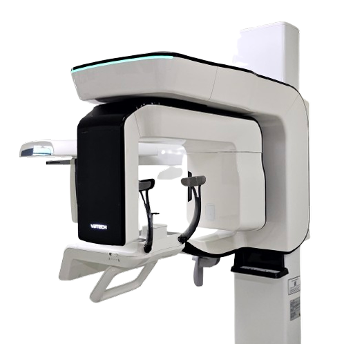
Model PaX-i3D SmartVatech
-
origin
Republic of Korea
-
Size(Capacity)
Dental Panorama, CBCT, and Cephalo
-
Features
2D AND 3D IN ONE VIEWER
PRODUCT DESCRIPTION
Dental X-rays can help your dentist detect oral health problems, such as cavities or gum disease, before they become serious. There are different types of dental X-rays, including intraoral (taken inside the mouth) and extraoral (taken outside the mouth). Dental X-rays are essential for proper oral health care.
CBCT(with Auto Pano), Panoramic, CBCT, and Cephallo
1 SCAN, 2 IMAGES
One scan with the Smart gives you not just a CT image but also an Auto Pano image. This means, patients who require both images do not need to undergo two X-ray scans. Also CT and Auto Pano images are displayed within the One Viewer feature.
2D AND 3D IN ONE VIEWER
Viewing 2D and 3D images together provides many benefits. There is no need to utilize two different software programs and it presents a professional look for your patients.
This layout helps patients better understand the images, which will eventually result in increasing acceptance rates
SMART Innovation for Accurate Diagnosis
- Anatomical FOV 12×9
- ART-V
ANATOMICAL FOV, 12X9
The innovative FOV of the PaX-i3D Smart provides an arch-shaped volume, which shows a wider view of dentition compared to other devices of the same FOV. Normally, a FOV 10×8.5 image shows tooth #8. However, when the tooth is lying on its side, there is a high possibility that the tooth will be cut out of the image. The “arch-shaped volume” eliminates this possibility and shows the hidden dentition area.
ART-V (ARTIFACT REDUCTION TECHNOLOGY OF VATECH)
The innovative FOV of the Vatech Paxi3D Smart provides an arch-shaped volume, which shows a wider view of dentition campared to other devices of the same FOV. Normally, a FOV 10×8.5 image shows tooth #8. However, when the tooth is lying on its side, there is a high possibility that the tooth will be cut out of the image. The “arch-shaped volume” eliminates this possibility and shows the hidden dentition area.
SAME WORDS, BUT DIFFERENT MEANING
Metal artifact hinders visualization and naturally reduces diagnostic confidence
PROFESSIONAL DIAGNOSTIC VALUE WITH PANORAMIC IMAGES
Smart Provides the most precise and high quality panoramic image. Clear and sharp panoramic image brings you better diagnostics. Enhanced details especially in the anterior and dental roots can be viewed. These consistently high quality images will become the new standard of panoramic imaging.
MAGIC PAN creates a superb panorama image. It is acquired through the elimination of distorted and blurred images caused by improper patient positioning(Optional). Focused image is reorganized throughout the whole dental arch and the image quality can be increased. The image becomes clearer especially in the area of incisor, canine , TMJ areas, and root canal.
PAYMENTS DETAILS
- Telegraphic Transfer : T/T
- Name : LEE, BYUNG-YOUNG
SHIPPING
- 56 Digital-ro 9-gil, Geumcheon-gu, Seoul (08512)
The person in charge
B.Y. LeeAddress
56 Digital-ro 9-gil, Geumcheon-gu, Seoul (08512)
-
- Business Type :
- Manufacturer
-
- Main Product :
- dental implant, dental X-ray
-
- Established :
- 2004-07-01
-
- Total Annual Revenue :
- 3~5 million (KRW)
-
- Total Employees :
- Less than 5
Please suggest a variety of your ideas such as design, impact, enhancements, etc
Please enter the text on the left image to prevent automatic input.
0 / 4000
CUSTOMER REVIEWS (0)

TRADE EXPERIENCE
-
- Total revenue
- 3~5 million (KRW)
-
- Total export revenue (previous year in USD)
- 350,000
-
- Number of foreign trade employees
- Less than 5
COMPARISON TO SIMILAR ITEMS more
- No Items
- supplier level
-
 PLATINUM
- ESSCO Limited Seller's Store
- Seller's Store url
- Response Level
★ ★ ★ ★ ★

- Supplier Level
★ ★ ★ ★ ★
- Transaction Level
★ ★ ★ ★ ★
- No Items


